Amethyst Piatra Neamt has officially opened its doors! Schedule an appointment
Breast cancer is the most common form of cancer among women in Romania. Every year, more than 8,000 women are diagnosed with breast cancer in Romania. Most (8 out of 10) are over 50, but younger women can also suffer from the condition.
A woman’s breasts are made up of fat, connective tissue and thousands of tiny glands, known as lobules, which produce milk. If a woman has a baby, milk is delivered to the nipple through small tubes called ducts, which allow her to breastfeed.
Our bodies are made up of billions of tiny cells. Normally, cells grow and multiply in an organised way. New cells appear only when they are needed. In cancer, this organised process works abnormally and cells start to grow and multiply in an uncontrolled way.

There are several different types of breast cancer, which can develop in different areas of the breast. Breast cancer is often classified into non-invasive and invasive types.
Non-invasive breast cancer is also known as cancer or carcinoma in situ, or pre-cancerous cells. This type of cancer is located in the ducts of the breast and has not developed the ability to spread outside the breast. This form of cancer rarely presents as a lump in the breast and is usually detected on a mammogram. The most common type of non-invasive cancer is ductal carcinoma in situ (DCIS).
Other less common types of breast cancer include invasive lobular breast cancer, which develops in the cells lining the milk-producing lobules, inflammatory breast cancer and Paget’s disease of the breast. Breast cancer can spread to other parts of the body, usually via the lymph nodes (small glands that filter bacteria from the body) or through the bloodstream. If extension occurs, the disease is known as secondary or metastatic breast cancer.
The first symptom of breast cancer that most women notice is a lump or area of thickened tissue in the breast. Most lumps (90%) are not cancerous, but it is always best to have a check-up with your GP or gynaecologist.
Please consult your family doctor or gynaecologist if you notice any of the following symptoms:
A change in the appearance of the nipple, as if sinking inside the breast, pain in either breast or in either armpit, unrelated to menstruation.
The causes of breast cancer are not fully understood. This means that it is difficult to say why one woman may develop breast cancer and another may not.
Certain issues, known as risk factors, can change the likelihood of someone developing breast cancer. There are some factors that cannot be done anything about, but other factors can be changed.
The risk of developing breast cancer increases with age. Breast cancer is more common in women over 50 who are past menopause. 8 out of 10 cases of breast cancer occur in women over 50.
For women between 50 and 70 years of age, it is advisable to have regular screening examinations for breast cancer..
If there are close relatives who have had breast cancer or ovarian cancer, you may have a higher risk of developing breast cancer. However, as breast cancer is the most common form of cancer in women, it is possible for it to occur more than once in the same family by chance.
Most cases of breast cancer are not hereditary (it is not passed on genetically), although studies have shown that certain genes, known as BRCA 1 and BRCA 2, can increase the risk of developing both breast and ovarian cancer. It is possible that these genes are passed from parent to child. A third gene (TP53) is also associated with increased risk of breast cancer.
If, for example, you have two or more close relatives on the same side of the family (e.g. mother, sister or daughter) who have had breast cancer at an age under 50, your risk of having the same disease is increased. That’s why it’s recommended that you have a genetic screening to check for those genes that make breast cancer likely. If you are concerned about your family history of breast cancer we advise you to discuss this with your GP.
If you have a history of breast cancer or incipient changes of non-invasive cancer cells in the breast ducts, you have a higher risk of developing cancer again in either the other breast or the same breast.
A benign breast lump does not mean you have breast cancer, but certain types of lumps can slightly increase your risk of developing it. Certain benign changes in breast tissue, such as atypical ductal hyperplasia (cells growing abnormally in the ducts) or lobular carcinoma in situ (abnormal cells in the breast lobes) increase the possibility of breast cancer.
The breasts are made up of thousands of small glands (lobules) that produce milk. This glandular tissue contains a higher concentration of breast cells compared to other breast tissue, making it denser. Women with denser breast tissue may have a higher risk of developing breast cancer because there are more cells that can become cancerous.
Dense breast tissue can make a breast scan (mammogram) more difficult to interpret because it makes any lumps or areas of abnormal tissue harder to detect. Younger women tend to have denser breasts. As we age, the amount of glandular tissue in the breasts decreases and is replaced by fat, so they become less dense.
In some cases, breast cancer cells can be stimulated to grow by estrogen, a female hormone. The ovaries, where the eggs are stored, start to produce estrogen when puberty sets in to regulate menstruation.
The risk of developing breast cancer can increase slightly depending on the amount of estrogen the body is exposed to. For example, if your first period was at a young age and menopause set in at an older age, you have been exposed to estrogen for a long time. Similarly, if you don’t have children, or if you had children later in life, your risk of developing breast cancer may increase slightly because your exposure to estrogen was not interrupted by pregnancy.
If you have gone through menopause and are overweight or obese, you are at higher risk of developing breast cancer. This is thought to be related to the amount of estrogen in your body, as being overweight or obese after menopause causes more estrogen to be produced.
Your risk of developing breast cancer may increase depending on how much alcohol you consume. Research shows that for every 200 women who typically consume two alcoholic drinks a day, there are three women with breast cancer compared to women who do not drink alcohol at all..
Certain medical procedures that use radiation, such as X-rays and CT (computed tomography) scans, can slightly increase the risk of developing breast cancer.
Hormone replacement therapy (HRT) is associated with a slightly increased risk of developing breast cancer. Both combined HRT and estrogen-only HRT therapy can increase the risk of developing breast cancer, although the risk is a little higher if you use combined HRT.
It is estimated that there would be an additional 19 cases of breast cancer for every 1,000 women taking combined HRT for 10 years. The risk continues to increase slightly with HRT, but returns to normal if you stop taking this type of therapy.
If your GP or gynaecologist has noticed typical breast cancer symptoms or if a screening mammogram has shown an abnormality, you will be asked to undergo further tests to confirm or refute the diagnosis of breast cancer.
If you have symptoms and have been referred by your GP or gynaecologist, you will have a mammogram, i.e. a breast X-ray. You may also need an ultrasound examination.
If you are under 35, your doctor may suggest that you just have an ultrasound. Younger women have denser breasts, which means mammography is not as effective as ultrasound for detecting cancer.
Ultrasound scans use high-frequency sound waves to produce an image that represents the internal structure of the breast. The image produced will show any lumps or abnormalities present in the breasts. Your doctor may also order an ultrasound if he or she needs to know if a lump is solid or if it contains fluid.
A biopsy involves taking a sample of breast tissue cells and testing it to see if the cells are cancerous. You may also need a scan and a puncture-biopsy of the lymph nodes in your armpit to see if they are also affected. Biopsies can be done in different ways and the type you have will depend on what your doctor knows about your condition. The different methods of conducting biopsies are outlined below.
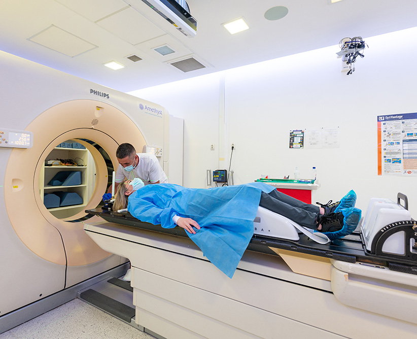
Computed Tomography (CT) scans, chest X-rays and liver ultrasound may be needed to check for extension of the cancer to the lungs or liver. An MRI of the breast may be needed to clarify or assess the extent of the disease within the breast.
If your doctor thinks there may be an extension of the cancer to your bones, you should have a bone scan. Before performing bone scintigraphy (a method of exploring organic tissue by introducing a radioactive substance into the body), a substance containing a small dose of radiation, known as an isotope, will be administered intravenously into the arm. If the bone is affected by cancer, this substance will be absorbed into the bone. Affected portions of bone will show up as intensely hyperfixed areas on the bone scan, which is performed using a special camera.
When breast cancer is diagnosed, doctors will give it a stage. Stage describes the size of the cancer and its extent.
It can provide accurate diagnostic information. T describes the size of the tumour, N describes whether the cancer has spread to the lymph nodes, and M gives an indication of whether the cancer has spread to other parts of the body.
The grade describes the appearance of the cancer cells.
People with breast cancer need to receive care from a multidisciplinary team, a team of specialists working together to provide the best treatment. They all form the Oncology Commission.
How to decide on the most effective treatment for breast cancer?
When deciding which treatment is best for you, doctors will consider the following:
• Usually, the first type of treatment for breast cancer is surgery. The type of surgery depends on the type of breast cancer you have. Surgery is usually followed by chemotherapy or radiotherapy or, in some cases, hormone treatments.
It should be remembered that the treatment you receive depends on your type of breast cancer. Your doctor will discuss the best treatment plan with you. Sometimes radiotherapy, chemotherapy or hormone therapy will be the first treatment option.
There are two types of surgery for breast cancer:
Studies have shown that breast-conserving surgery followed by radiotherapy is as useful as total mastectomy in the treatment of early-stage breast cancer.
Conservative surgery ranges from a lumpectomy to wide local excision, in which only the tumour and a small amount of adjacent tissue is removed, to a partial mastectomy or sectoral excision, in which up to a quarter of the breast is removed.
If you are having breast-conserving surgery, the amount of breast tissue that will be removed will depend on:
Your surgeon will always remove an area of healthy breast tissue around the cancer, which will be tested to see if there is any sign of cancer. If there are no signs of cancer in healthy tissue, there is less chance of recurrence. If cancer cells are found in the adjacent tissue, additional breast tissue may need to be removed.
Usually, after breast-conserving surgery, you will be offered the option of undergoing radiotherapy treatment to destroy the remaining cancer cells.
Mastectomy is the removal of all breast tissue, including the nipple. If there are no definite signs that the cancer has spread to the lymph nodes, you may need to have a mastectomy, a procedure that removes your entire breast and biopsies a sentinel lymph node (SLNB).
If cancer has spread to the lymph nodes, extensive removal (removal) of lymph nodes in the axilla (armpit area) may be needed.
Breast reconstruction is surgery used to reshape the breast to look as much like the other breast as possible. Reconstruction can be performed at the same time as the mastectomy (immediate reconstruction) or it can be performed later (delayed reconstruction). This can be done either by breast implant or by using your own tissue from another part of the body to create a new breast.
To see if the cancer has spread, a procedure called a sentinel lymph node biopsy (SLNB) may be performed. Sentinel lymph nodes are the first lymph nodes affected by cancer cells if they spread. These are part of the lymph nodes in the underarm area (axillary lymph nodes). The position of sentinel lymph nodes varies, so they are identified using a combination of radioisotope and a blue contrast agent.
Sentinel lymph nodes are examined in the laboratory to see if they have cancer cells. This procedure is a good indicator of whether or not the cancer has spread.
If there are cancer cells in the sentinel lymph nodes, another surgery may be needed to remove more lymph nodes from the axillary region.
You can follow one or a combination of these treatments. The type of treatment or combination of treatments will depend on how the cancer was diagnosed and what stage it is in. Breast cancer diagnosed at screening may be at an early stage, but breast cancer diagnosed when you have symptoms may be at a more advanced stage and require different treatment. The Oncology Commission will discuss the most appropriate treatments and let you know your options.
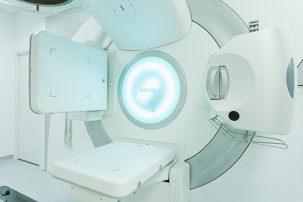
Radiotherapy uses controlled doses of radiation to destroy cancer cells. This can be done before surgery to reduce the size of the tumour or after surgery and chemotherapy to destroy the remaining cancer cells. Sometimes it is sufficient to use it as an exclusive and curative treatment.
Studies show that radiotherapy after a conservative mastectomy significantly increases patients’ chances of survival.
With the latest IMRT technology, and even more so with the latest VMAT technology, there is now the possibility to protect organs at risk that are close to the tumour from being affected by the treatment itself.
If you need post-operative radiotherapy, your treatment will start in 1-2 months after surgery or chemotherapy to give your body a chance to recover. You will most likely need radiotherapy sessions five times a week, for a period of three to six weeks. Each session will last only a few minutes.
The type of radiotherapy you have depends on the type of cancer you have.
Possible side effects of radiotherapy include:
Chemotherapy involves the use of anti-cancer (cytotoxic) drugs to destroy cancer cells. Chemotherapy is usually used after surgery to destroy cancer cells that have not been removed. This is called adjuvant chemotherapy. In some cases chemotherapy may be used before surgery, which is generally used to shrink a larger tumour. This is called neo-adjuvant chemotherapy. Several different drugs are used for chemotherapy, often in combination. The choice of drugs and combination depends on the type of breast cancer and how far it has spread.
Chemotherapy is usually offered as an outpatient treatment, which means there is no need to stay in hospital overnight. As a rule, the drugs are administered in a vein by drip directly into the bloodstream. In some cases, you may be given tablets that you can take at home. You may receive chemotherapy sessions every two or three weeks for four to eight months to give your body a break between treatments.
The main side effects of chemotherapy include:
These can be prevented or controlled with medicines that your doctor can prescribe.
Chemotherapy drugs can also stop the body’s production of estrogen. Estrogen is known to promote the growth of certain types of breast cancers. If you have not gone through menopause, your periods may stop while you are undergoing chemotherapy treatment. After you have finished chemotherapy, your ovaries may produce estrogen again. However, in some cases this does not happen and menopause will set in early. This is more likely to happen in women over 40, closer to menopausal age.
Your doctor will discuss with you the impact that the treatment will have on your fertility.
You can follow one or a combination of these treatments. The type of treatment or combination of treatments will depend on how the cancer was diagnosed and what stage it is in. Breast cancer diagnosed at screening may be at an early stage, but breast cancer diagnosed when you have symptoms may be at a more advanced stage and require different treatment. The Oncology Commission will discuss the most appropriate treatments and let you know your options.